

Summary video
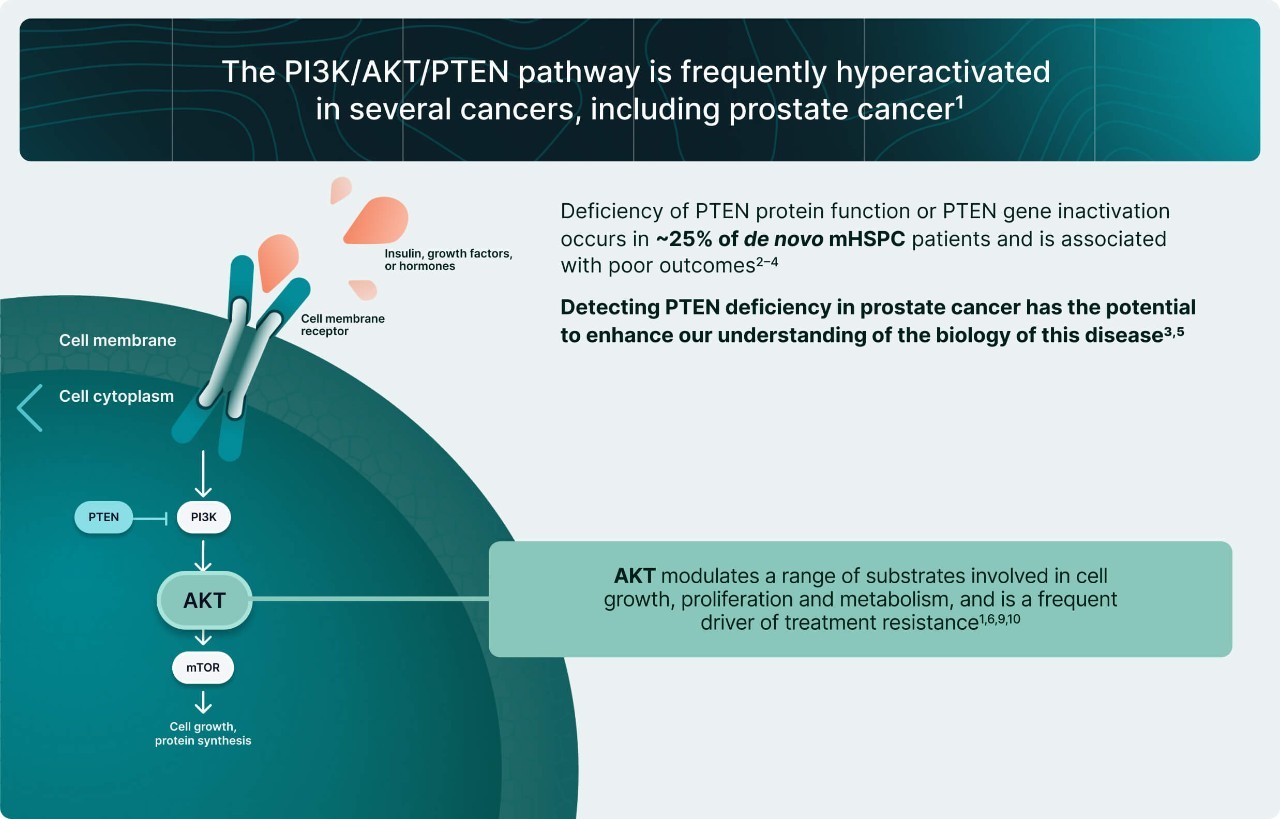
The PI3K/AKT pathway is the most frequently disrupted signaling pathway in human cancers
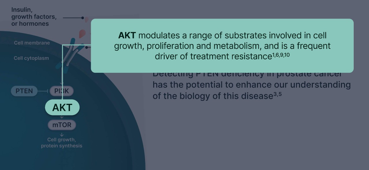
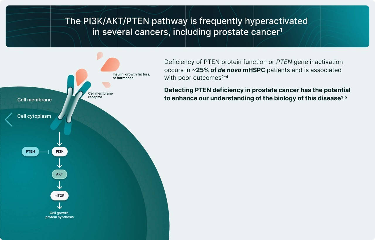
AKT plays a central role in the pathway, modulating a range of substrates to promote cell growth, proliferation, metabolism and survival.1
In prostate cancer, overactivation of the PI3K/AKT pathway contributes to1:
- Tumor growth
- Disease progression
- Development of castration resistance
50–70%
Proportion of metastatic prostate
cancers with PI3K/AKT pathway overactivation.2,3
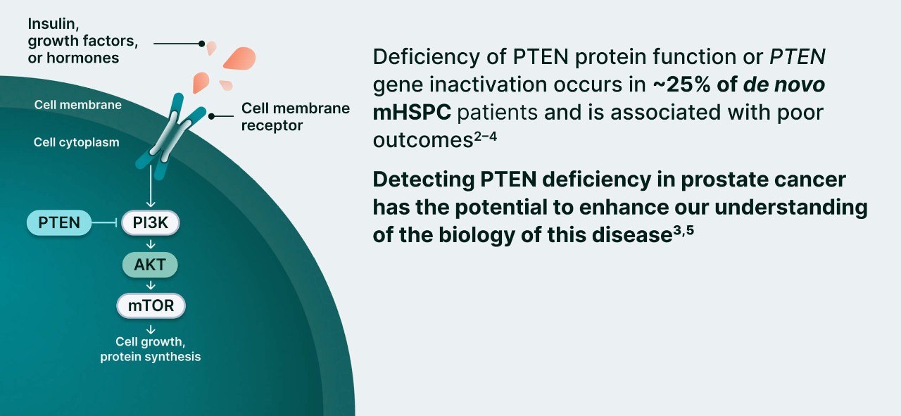
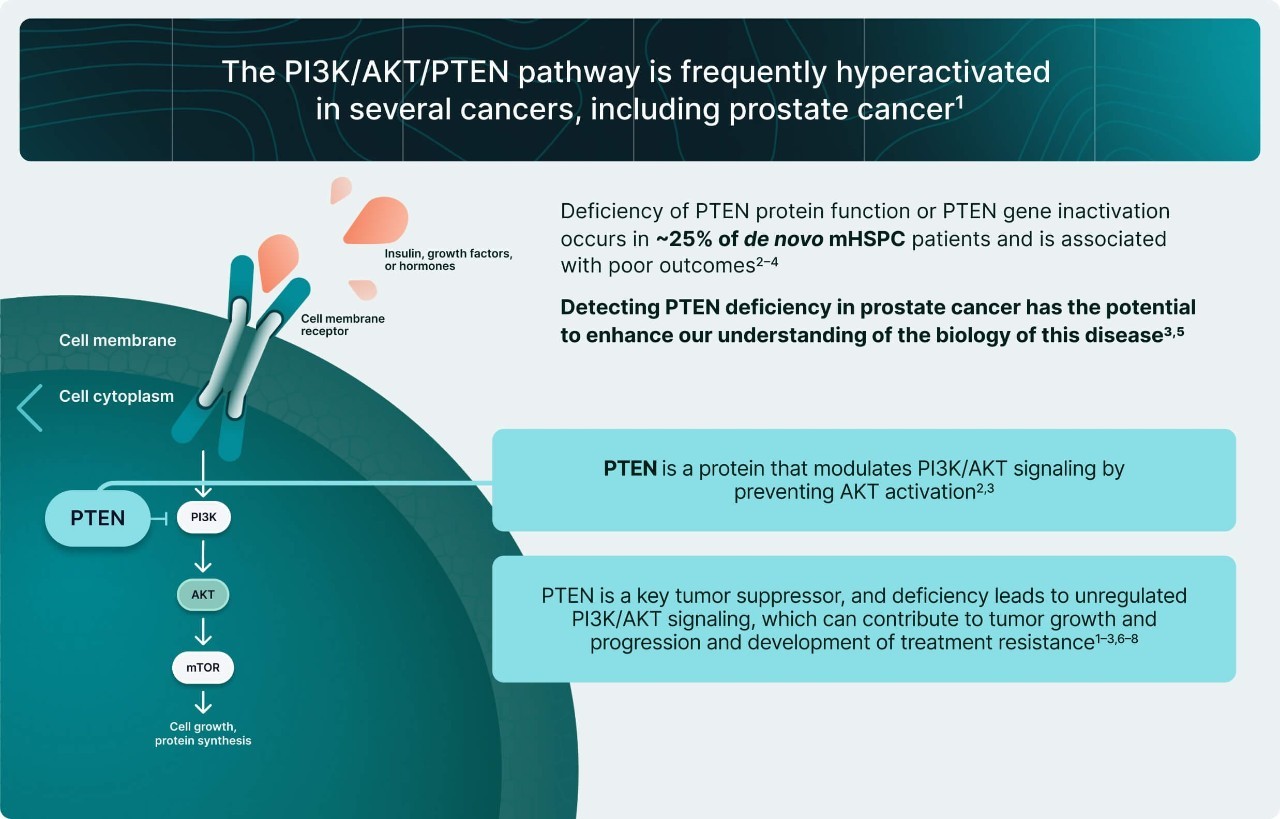
PTEN deficiency is one of the main causes of PI3K/AKT pathway overactivation in prostate cancers
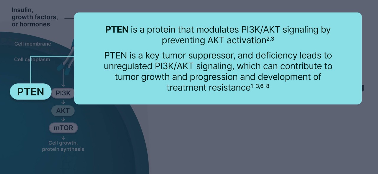
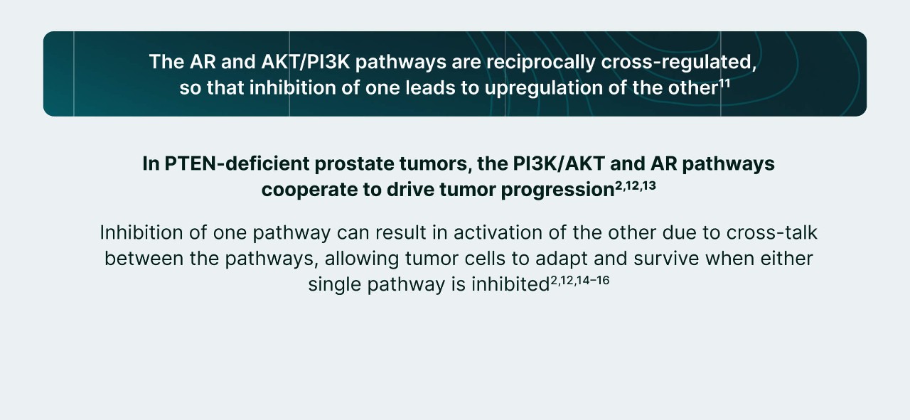
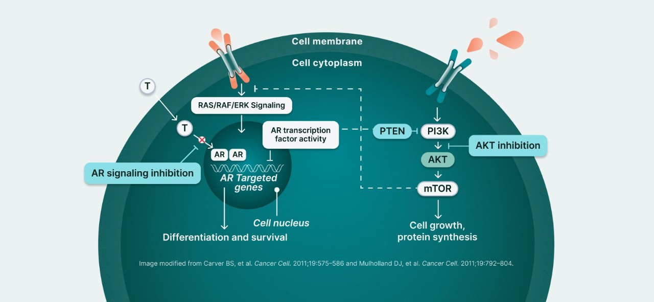
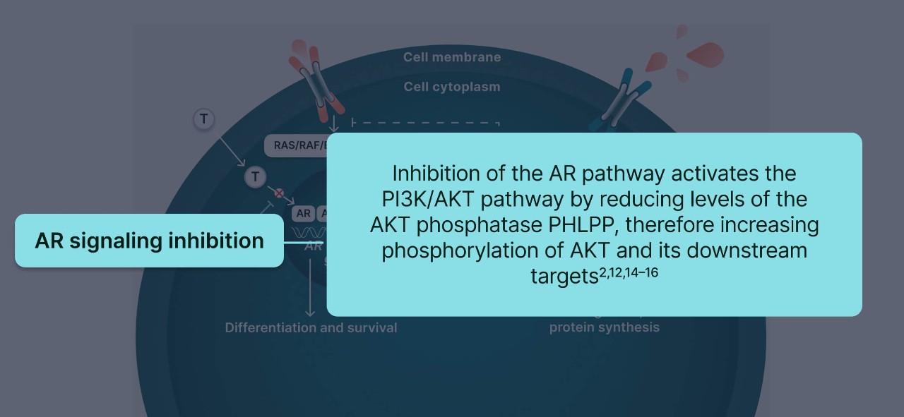
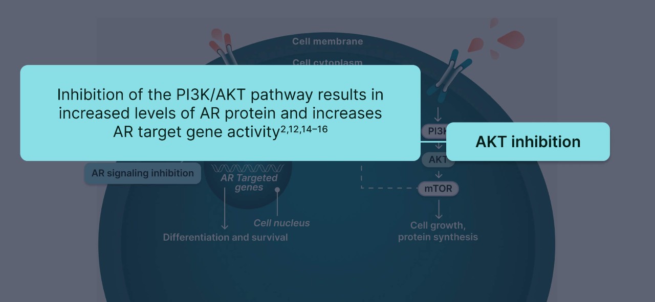
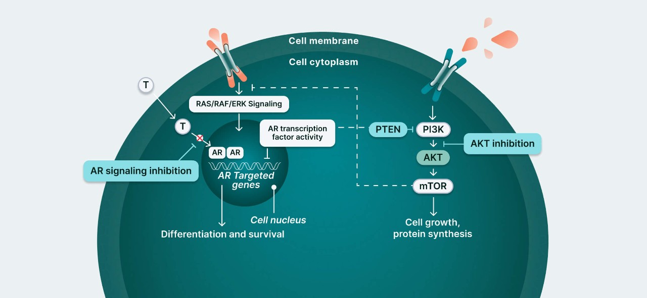
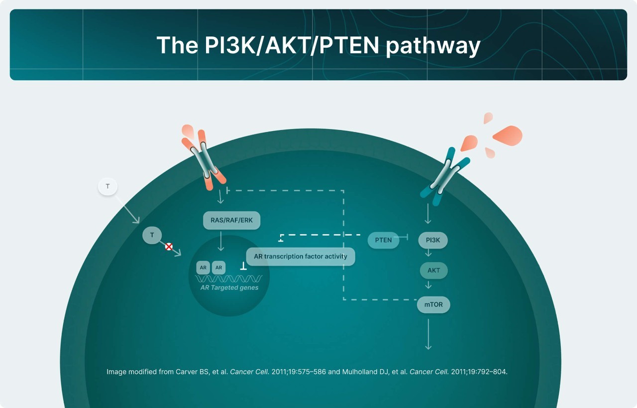
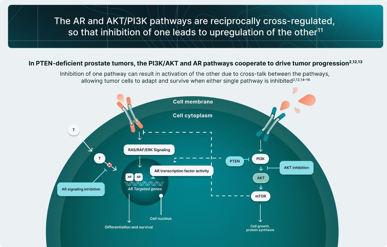
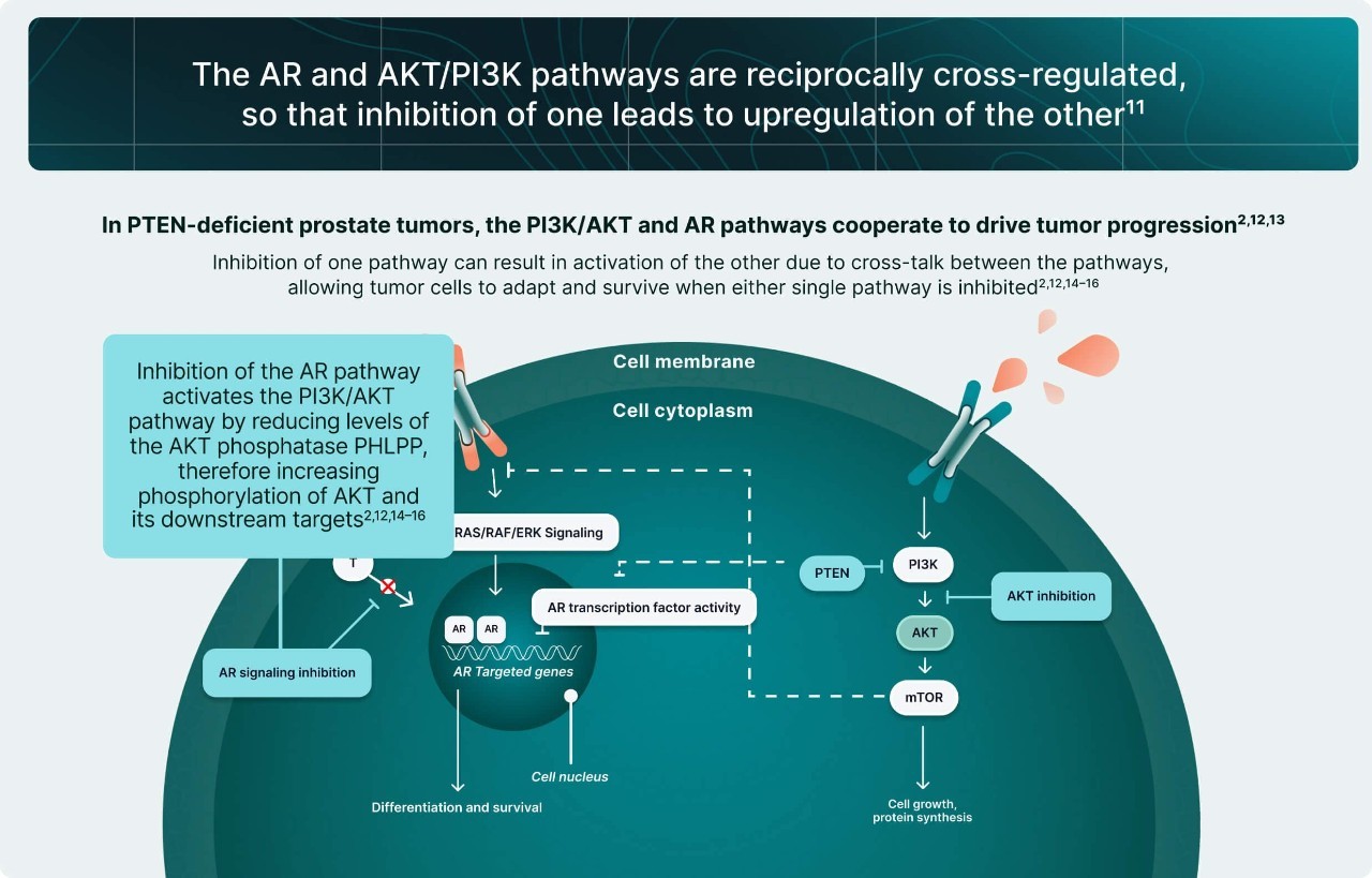
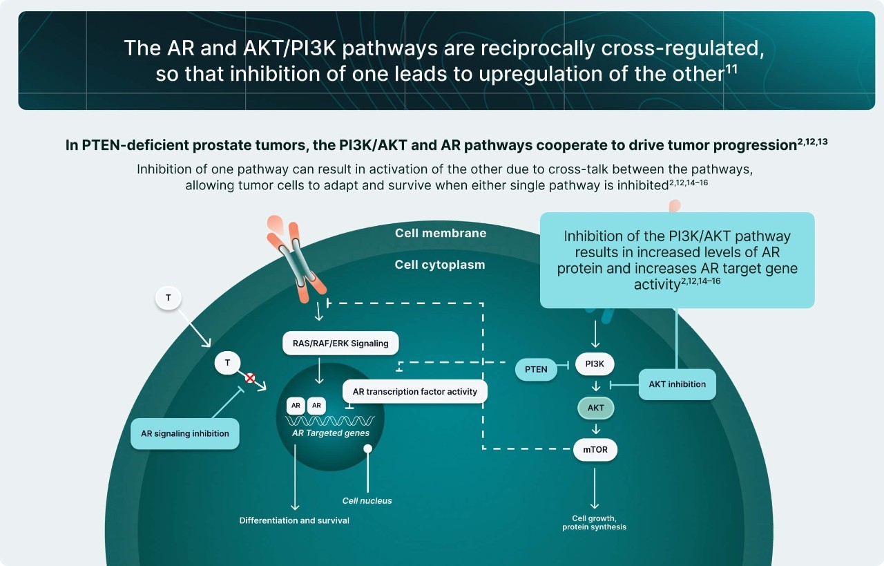
PTEN is a key tumor suppressor protein that modulates the PI3K/AKT pathway by preventing AKT activation.1,4 In PTEN-deficient prostate tumors, the PI3K/AKT pathway provides an additional proliferative drive that complements AR signaling, promoting tumor progression and castration resistance.1,3,5
Uncover more about the clinical implications of PI3K/AKT pathway activation and its interaction with AR signaling in PTEN-deficient tumors.


-
rotate-slide
-
slide-1
-
slide-2
-
slide-3
-
slide-4
-
slide-5
-
slide-6
-
slide-7
-
slide-7
-
slide-8
-
slide-1
-
slide-2
-
slide-3
-
slide-4
-
slide-5
-
slide-6
-
slide-7
-
slide-8
Detecting PTEN deficiency has potential to enhance understanding of drivers of disease progression
Knowing a patient’s PTEN status can inform understanding of whether PI3K/AKT pathway activation is driving a patient’s disease progression and castration resistance.6
Ongoing research is further investigating the PI3K/AKT pathway and PTEN deficiency in metastatic prostate cancer, with AKT representing an ideal therapeutic target due to its central role in the pathway.1,6
knowledge check
AKT, protein kinase B; AR, androgen receptor; PI3K, phosphoinositide 3-kinase; PTEN, phosphatase and tensin homolog.
1. Brown JS, Banerji U. Pharmacol Ther. 2017;172:101–115; 2. Ikeda S, et al. Cancer Biol Ther. 2019;20(2):219–226; 3. Carver BS, et al. Cancer Cell. 2011;19(5):575–586; 4. Jamaspishvili T, et al. Nat Rev Urol. 2018;15(4):222–234;2018; 15(4):222–234; 5. Mulholland DJ, et al. Cancer Cell. 2011;19(6):792–804; 6. Turnham DJ, et al. Cells. 2020;9(11):2342.