

Summary video
Biomarkers are key to precision oncology
Biomarkers provide important clinical information to help stratify patients in terms of4:

Diagnosis

Prognosis

Response to therapies
It is very exciting to be a urologist today, seeing how biomarkers are proving to be invaluable tools in the diagnosis, prognosis and treatment of prostate cancer.

Biological pathways are altered in metastatic prostate tumors
Metastatic prostate tumors are often characterized by gene alterations in several biological pathways, including5–7:
- Androgen receptor (AR) pathway
- PI3K/AKT/PTEN signaling pathway
- Wnt pathway
- Cell cycle and DNA repair pathways
In metastatic prostate cancer, genomic and protein biomarkers are an increasingly important tool to better understand patient prognosis and risk of disease progression, and to inform personalized treatment selection.1,4
Discover the PI3K/AKT/PTEN signaling pathway
The PI3K/AKT pathway is the most frequently disrupted signaling pathway in human cancers.8
In metastatic prostate cancer, PI3K/AKT pathway overactivation occurs in 50–70% of cases, and is often the result of a deficiency in the key tumor suppressor protein, PTEN.5,9
Explore the pathway
Guidelines recommend biomarker testing of metastatic prostate tumors
Both germline testing and somatic tumor testing are recommended for metastatic prostate tumors (mHSPC and mCRPC), with re-evaluation of the tumor molecular profile through repeat somatic testing at the time of disease progression.1,2
Somatic tumor testing of metastatic prostate tumors can identify biomarkers, such as HRR alterations, MSI-H or dMMR, which can provide prognostic information as well as inform treatment decision-making, including eligibility for biomarker-driven therapies.1–3
Testing is recommended by key medical oncology and urology guidelines, including1–3:
- National Comprehensive Cancer Network® (NCCN®)
- American Society of Clinical Oncology
- American Urological Association/Society of Urologic Oncology
Biomarker testing workflow: Clinical guidelines
Stay at the forefront of biomarker testing for metastatic prostate cancer with a simplified, step-by-step workflow integrating key recommendations from clinical guidelines.
View and download (PDF)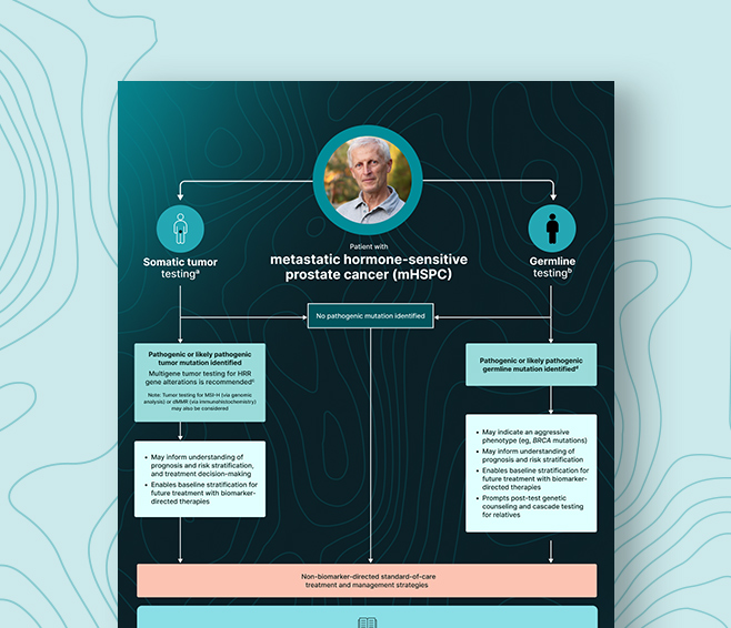
It is our obligation to identify patients who may be eligible for precision-based therapies. Failing to test for appropriate biomarkers could result in lost opportunities for significantly improved outcomes.

Ongoing research: The value of biomarker-driven approaches earlier in the patient journey
Although there are no biomarker-driven therapies approved for mHSPC, ongoing research is exploring the potential for personalized approaches to help improve outcomes earlier in the disease, before progression to mCRPC.2,10–12
As the clinical landscape continues to shift toward precision medicine, improved understanding of biomarker-driven approaches may help to refine targeted strategies for patients with mHSPC.12,13
knowledge check
AKT, protein kinase B; AR, androgen receptor; dMMR, DNA mismatch repair; DNA, deoxyribonucleic acid; HRR, homologous recombination repair; mCRPC, metastatic castration-resistant prostate cancer; mHSPC, metastatic hormone-sensitive prostate cancer; MSI-H, microsatellite instability-high; PI3K, phosphoinositide 3-kinase; PTEN, phosphatase and tensin homolog; Wnt, wingless-type.
1. Referenced with permission from the NCCN Clinical Practice Guidelines in Oncology (NCCN Guidelines®) for Prostate Cancer V.2.2026. © National Comprehensive Cancer Network, Inc. 2026. All rights reserved. Accessed September 15, 2025. To view the most recent and complete version of the guideline, go online to NCCN.org. NCCN makes no warranties of any kind whatsoever regarding their content, use or application and disclaims any responsibility for their application or use in any way; 2. Yu EY, et al. J Clin Oncol. 2025;43(6):748–758; 3. Lowrance W, et al. J Urol. 2023;209(6):1082–1090; 4. Pulido R, et al. Cold Spring Harb Perspect Med. 2019;9(12):a036293; 5. Ikeda S, et al. Cancer Biol Ther. 2019;20(2):219–226; 6. Robinson D, et al. Cell. 2015;161(5):215–228; 7. Wasim S, et al. Int J Mol Sci. 2022;23(22):14257; 8. Brown JS, Banerji U. Pharmacol Ther. 2017;172:101–115; 9. Carver BS, et al. Cancer Cell. 2011;19(5):575–586; 10. Ng K, et al. Oncol Ther. 2020;8(2):209–230; 11. Chen K, et al. World J Urol. 2023;41(8):2021–2031; 12. Meagher MF, et al. Res Rep Urol. 2023;15:509–517; 13. Turnham DJ, et al. Cells. 2020;9(11):2342.